Copyright Southeastern Orthopaedic Specialists, P.A. All rights reserved.
Read our Legal Disclaimer. Web development by A Better Web, Inc.
Radiography (X-Rays)
X-rays (radiographs) are the most common and widely available diagnostic imaging technique. Even if you just complain about a sprain in your wrist or ankle, your doctor will probably order radiographs to make sure that no bone is broken. X-rays are always used for fractures and joint dislocations, and may also be recommended if your doctor suspects damage to a bone or joint from other conditions such as arthritis or osteonecrosis (bone cell death). The part of your body being pictured is positioned between the X-ray machine and photographic film. As you hold still, the machine briefly sends electromagnetic waves (radiation) through your body. This exposes the film, creating a picture of your internal structure. The level of radiation exposure from X-rays is minimal, but your doctor will take special precautions if you are pregnant. Bones, tumors and other dense matter appear white or light because they absorb the radiation. Soft tissues and breaks in bone let radiation pass through, making these parts look darker. Sometimes, to make certain organs stand out in the picture, you are asked to drink barium sulfate or be injected with a dye. Several X-rays from different angles may be needed. If you have a fracture in one limb, your doctor may want a comparison X-ray of your uninjured limb. Your X-ray session will probably take 10 to 15 minutes; no specific preparations are required. Each Division of Southeastern Orthopaedic Specialists, P.A. is equipped with state of the art X-ray equipment and facilities. Our facilities utilize high tech digital technology as well as traditional film technology for special views. Our offices have the capability to send your X-ray electronically (and privately) to your physician’s work station or to your exam room. We can also instantly send your “digital films” to those physicians whom might assist you doctor in their interpretation, such as at the hospital or in private practice. But be assured that you confidentiality is maintained at all times.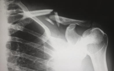


![[x]](index_html_files/close.png "Close")

Radiography (X-Rays)
X-rays (radiographs) are the most common and widely available diagnostic imaging technique. Even if you just complain about a sprain in your wrist or ankle, your doctor will probably order radiographs to make sure that no bone is broken. X-rays are always used for fractures and joint dislocations, and may also be recommended if your doctor suspects damage to a bone or joint from other conditions such as arthritis or osteonecrosis (bone cell death). The part of your body being pictured is positioned between the X-ray machine and photographic film. As you hold still, the machine briefly sends electromagnetic waves (radiation) through your body. This exposes the film, creating a picture of your internal structure. The level of radiation exposure from X-rays is minimal, but your doctor will take special precautions if you are pregnant. Bones, tumors and other dense matter appear white or light because they absorb the radiation. Soft tissues and breaks in bone let radiation pass through, making these parts look darker. Sometimes, to make certain organs stand out in the picture, you are asked to drink barium sulfate or be injected with a dye. Several X-rays from different angles may be needed. If you have a fracture in one limb, your doctor may want a comparison X-ray of your uninjured limb. Your X-ray session will probably take 10 to 15 minutes; no specific preparations are required. Each Division of Southeastern Orthopaedic Specialists, P.A. is equipped with state of the art X-ray equipment and facilities. Our facilities utilize high tech digital technology as well as traditional film technology for special views. Our offices have the capability to send your X-ray electronically (and privately) to your physician’s work station or to your exam room. We can also instantly send your “digital films” to those physicians whom might assist you doctor in their interpretation, such as at the hospital or in private practice. But be assured that you confidentiality is maintained at all times.
Copyright
Southeastern Orthopaedic Specialists, P.A.
All rights reserved | Read our Legal Disclaimer
Web development by A Better Web, Inc.