Copyright Southeastern Orthopaedic Specialists, P.A. All rights reserved.
Read our Legal Disclaimer. Web development by A Better Web, Inc.
Electromyography (EMG)
An electromyography (EMG) records and analyzes the electrical activity in your muscles. It is used to learn more about the functioning of nerves in the arms and legs. For example, a fracture of the upper arm bone (humerus) may tear or pinch the radial nerve. An EMG can be used to identify the damage if nerve function doesn't return within 4 months of the injury. During an EMG, small, thin needles are placed in the muscle to record the electrical activity. When the needles are inserted, you may feel some pain and discomfort. The doctor will ask you to relax the muscle and then to tense it slightly. The electrical signals generated by your muscle are broadcast on a TV-like screen. When the needles are removed, you may experience some soreness and bruising, but this will disappear in a few days. There are no long-term side effects. If you are taking blood-thinning medications, have lung disease or are at risk for infection, tell the physician who is conducting the test. On the day of the test, do not put any lotions or creams on the area to be tested and do not wear any jewelry. Usually, you can get the results immediately after the test. All Divisions of Southeastern Orthopaedic Specialists, P.A. have Board Certified Physical Medicine and Rehabilitation Physicians who are Interventional Specialists trained in the administration and interpretation of these studies.
Image courtesy of ViewMedica


![[x]](index_html_files/close.png "Close")

Electromyography (EMG)
An electromyography (EMG) records and analyzes the electrical activity in your muscles. It is used to learn more about the functioning of nerves in the arms and legs. For example, a fracture of the upper arm bone (humerus) may tear or pinch the radial nerve. An EMG can be used to identify the damage if nerve function doesn't return within 4 months of the injury. During an EMG, small, thin needles are placed in the muscle to record the electrical activity. When the needles are inserted, you may feel some pain and discomfort. The doctor will ask you to relax the muscle and then to tense it slightly. The electrical signals generated by your muscle are broadcast on a TV-like screen. When the needles are removed, you may experience some soreness and bruising, but this will disappear in a few days. There are no long-term side effects. If you are taking blood-thinning medications, have lung disease or are at risk for infection, tell the physician who is conducting the test. On the day of the test, do not put any lotions or creams on the area to be tested and do not wear any jewelry. Usually, you can get the results immediately after the test. All Divisions of Southeastern Orthopaedic Specialists, P.A. have Board Certified Physical Medicine and Rehabilitation Physicians who are Interventional Specialists trained in the administration and interpretation of these studies.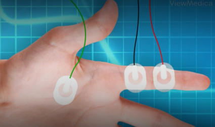
Copyright
Southeastern Orthopaedic Specialists, P.A.
All rights reserved | Read our Legal Disclaimer
Web development by A Better Web, Inc.